Clinical Catalogue & Surgical Portfolio

Orthopaedic Surgical Specialties & Joint Care

Evidence-based reconstructive procedures, tissue-sparing approaches, robotic-assisted and patient-specific joint replacement, complex revision hip and knee surgery, and advanced knee ligament reconstruction.

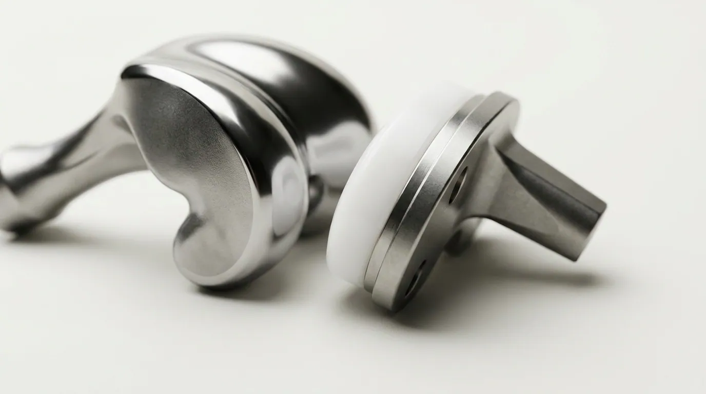
Precision Total Knee Replacement Implant
Sub-Millimeter BalanceKinematic Fit

Specialty Intervention 02 · Knee Joint

Total Knee Replacement

Moving past rigid mechanical alignment, Dr. Ya'ish implements kinematic and patient-specific protocols calibrated to replicate each patient's constitutional joint line and soft-tissue envelope. This physiological philosophy reduces unnatural tightness and restores a natural, pain-free arc of flexion.

Alignment Philosophy

Kinematic Restoration

Component Fixation

Cobalt-Chrome & PEEK/Poly

Soft Tissue Release

Preservative / Negligible

Functional Target

125°+ Smooth Flexion

Digital Pre-Op Templating
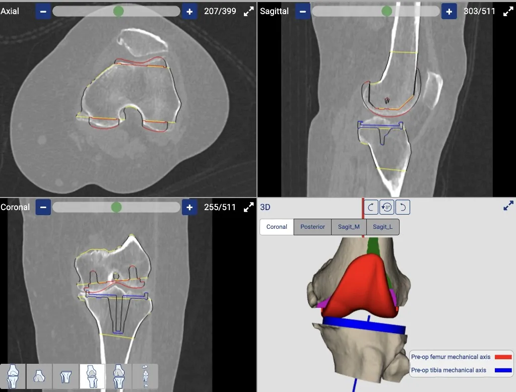
CT-based patient-specific knee replacement planning in axial, sagittal, coronal and 3D views
CT PlanningPatient-Specific Guides

Specialty Intervention · Personalised Knee Planning

Patient-Specific Knee Replacement (PSI)

CT-based pre-operative planning produces patient-specific instrumentation, so femoral and tibial cuts are mapped to the individual's own anatomy before the first incision is made. PSI sits alongside total knee replacement; it is the planning method, not a different operation.

Procedure

Total knee replacement with PSI

Planning

CT before the first incision

Instrumentation

Guides matched to the patient's anatomy

Role

Alongside total knee replacement

PSI sits alongside total knee replacement

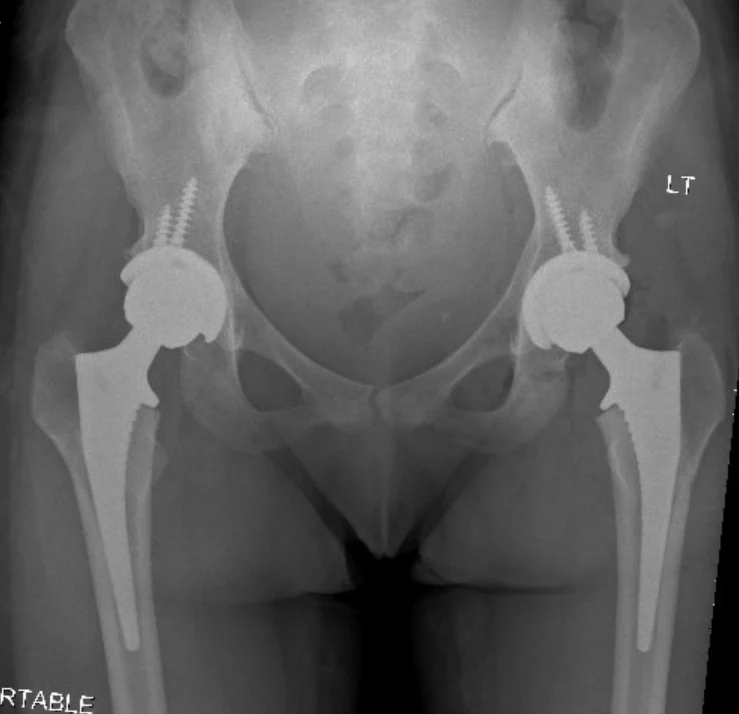
Radiograph showing bilateral total hip replacements
Cementless or HybridLeg-Length Restoration

Specialty Intervention · Hip Joint

Hip Replacement

Primary total hip arthroplasty using cementless and hybrid fixation, with leg-length and offset restored to the patient's own anatomy.

Procedure

Primary Total Hip Arthroplasty

Fixation

Cementless or Hybrid

Anatomic Target

Leg length and offset

Planning

CT templating when indicated

Primary hip replacement

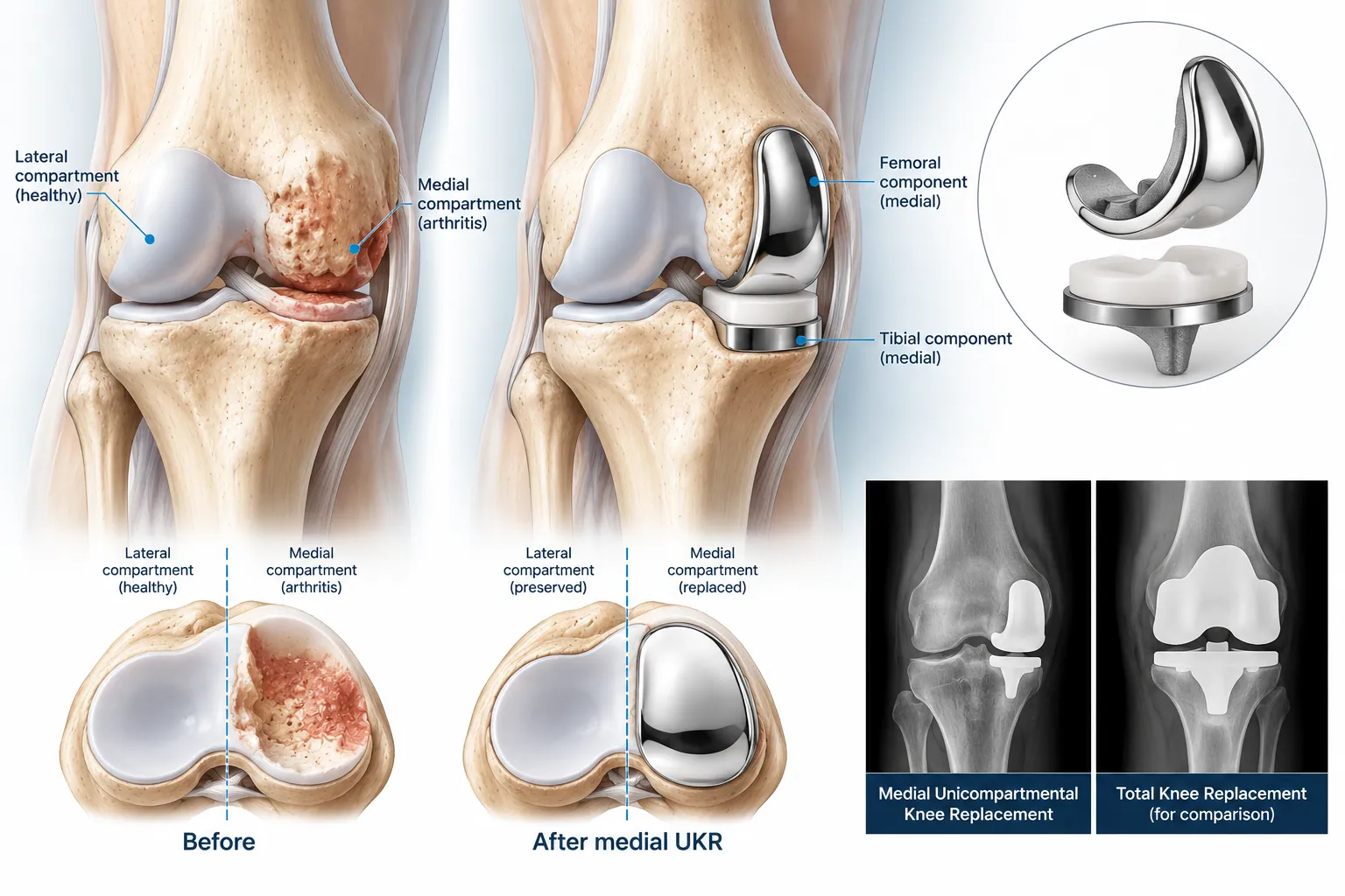
Anatomical comparison of a knee with medial compartment arthritis and the same knee after partial knee replacement
Cruciate-RetainingOutpatient Eligible

Specialty Intervention 03 · Compartmental Resurfacing

Partial Knee Replacement

Ideal for isolated medial or lateral compartment osteoarthritis. By resurfacing solely the worn articulating compartment, both the Anterior Cruciate Ligament (ACL) and Posterior Cruciate Ligament (PCL) remain fully intact, preserving proprioceptive feedback and a more natural knee. Quicker recovery to function representing native joint.

Native Ligaments

100% Retained

Incision Length

6 - 8 cm Micro-Incised

Hospital Stay

Day-Case / Overnight

Normal Activity

3 - 4 Weeks

Medial & Patellofemoral

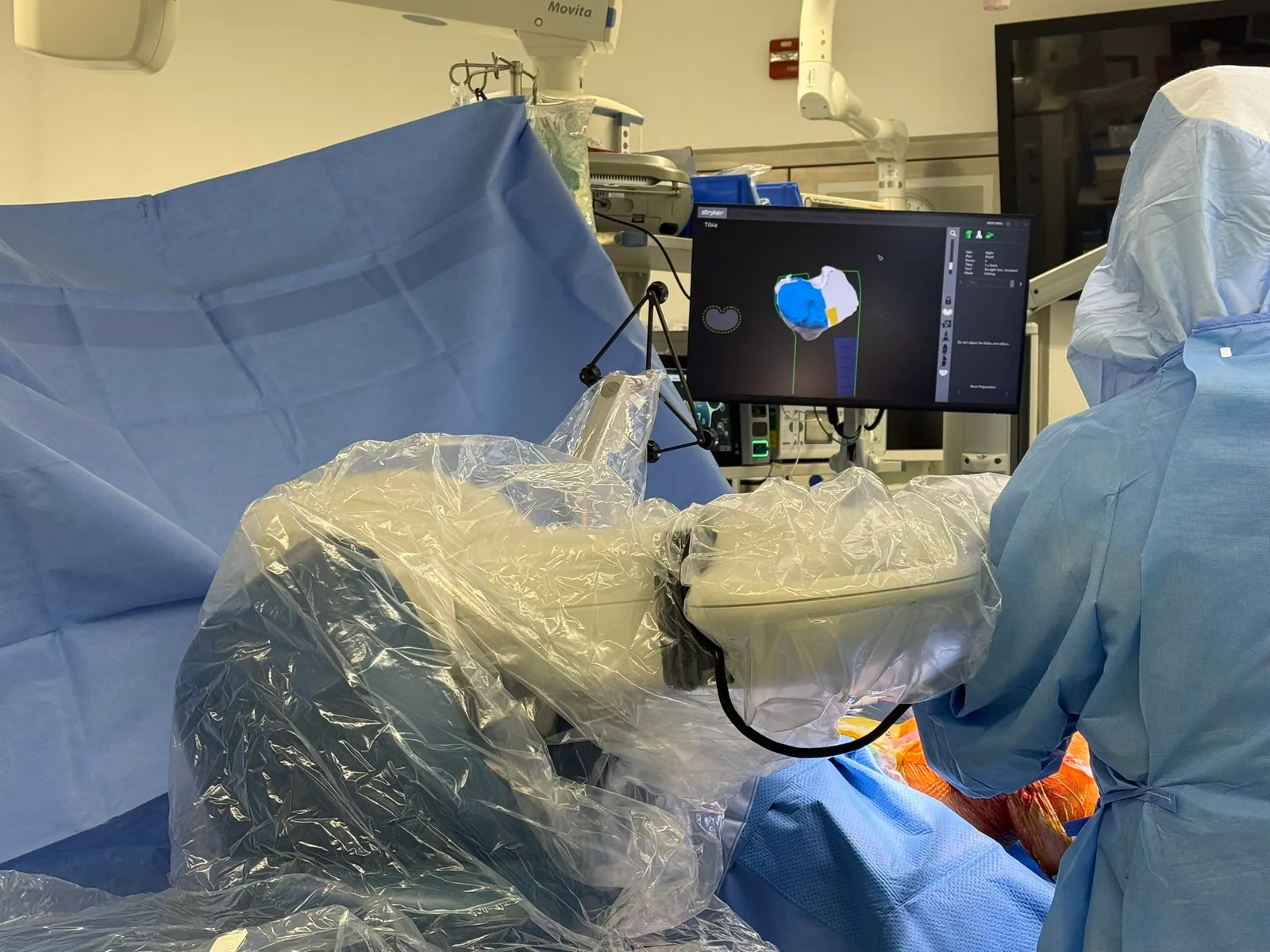
Stryker Mako robotic arm positioned in theatre with the intra-operative navigation display
CT 3D ModelingHaptic Security

Specialty Intervention 04 · State-of-the-Art Navigation

Robotic Joint Replacement

Next-generation robotic arm stereotactic guidance combined with high-resolution pre-operative 3D-CT modeling. Surgeon-controlled haptic boundaries prevent inadvertent soft-tissue damage, keeping femoral and tibial component positioning within 0.5 mm and 0.5 degrees of planned alignment.

Positional Precision

±0.5 mm / 0.5°

Real-Time Balancing

Dynamic Ligament Sensor

Soft Tissue Safety

Haptic Boundary Lock

Revision Risk Reduction

Clinically Documented

Robotic planning when indicated

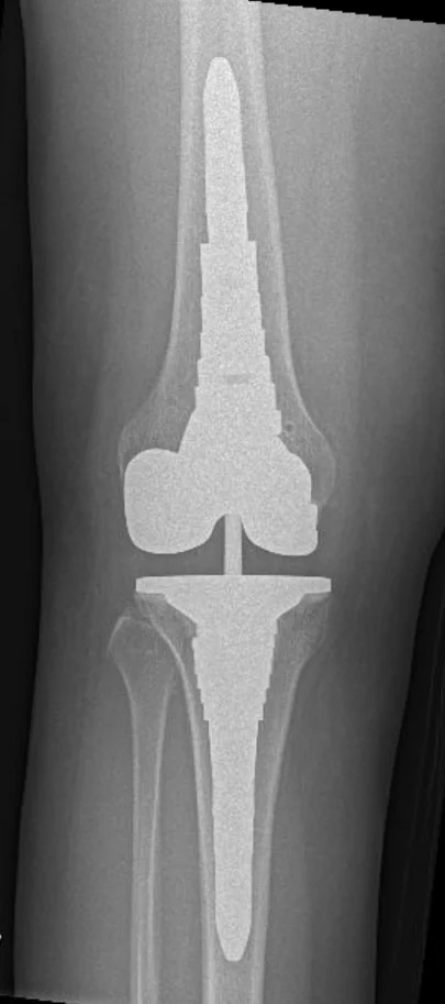
Radiograph of a revision total knee replacement with long-stem femoral and tibial components
Tertiary Level ReferralAO Recon Expert

Specialty Intervention 05 · High-Complexity Reconstruction

Revision Hip & Knee Surgery

Specialized referral pathway for previously operated joints with aseptic loosening, osteolysis, structural bone loss, instability, or periprosthetic joint infection (PJI). Uses modular revision stems, titanium trabecular 3D cones, custom acetabular augments, and hinge knee systems.

Indications Managed

Loosening, PJI, Bone Loss

Bone Reconstruction

Trabecular Titanium Sleeves

MDT Collaboration

Infectious Disease & Radiology

Regional Expertise

AO Recon MENA Faculty

Major Referral Center

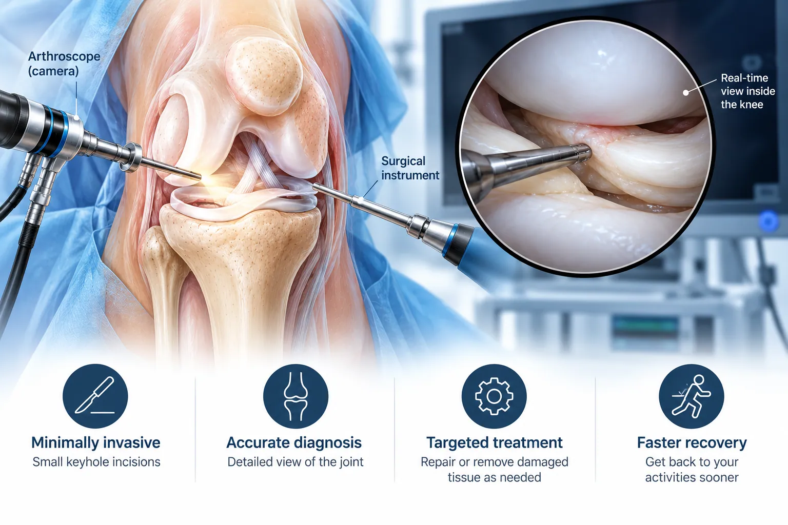
Knee arthroscopy showing the arthroscope camera and surgical instrument with the real-time view inside the joint
Keyhole SurgeryMeniscal Salvage

Specialty Intervention 06 · Knee Sports Injuries

Knee Arthroscopy

Minimally invasive keyhole surgery for meniscal tears, cartilage lesions, and mechanical locking of the knee. High-definition arthroscopy allows targeted repair or debridement while protecting healthy tissue.

Procedures

Meniscus / Cartilage

Meniscal Strategy

Preservation & All-Inside Fix

Surgical Access

4 mm HD Arthroscopy

Recovery

Repair-specific protocol

Repair-first meniscal strategy

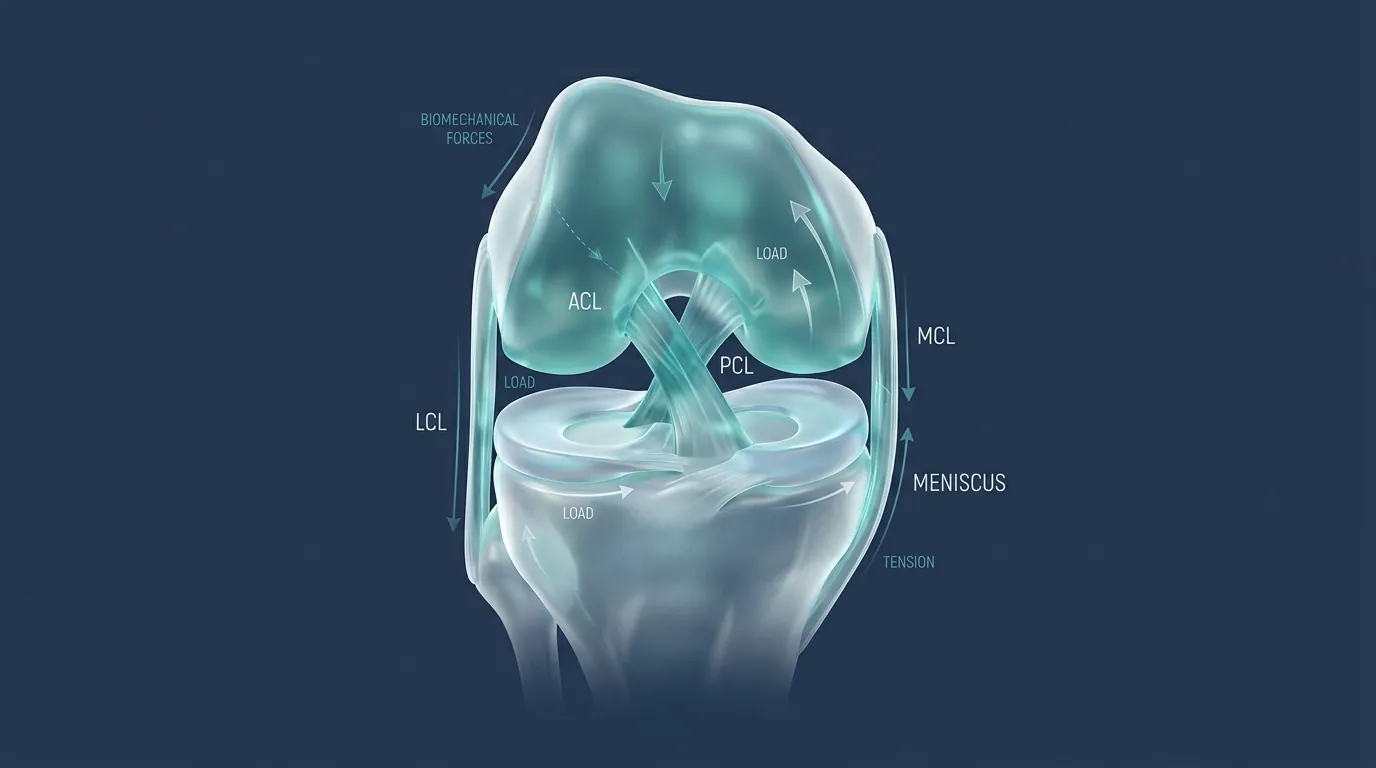
Knee ligament anatomy including the ACL
ACL ReconstructionAnatomic Kinematics

Specialty Intervention 07 · Knee Sports Injuries

Knee Ligament Reconstruction, including ACL

Anatomical reconstruction of the anterior cruciate ligament (ACL) and associated knee ligaments after rupture or multi-ligament injury. The goal is a stable, proprioceptive knee for daily function, prayer, work, and return to sport when appropriate.

Procedures

ACL / PCL / ALL / MPFL

Focus

Knee ligament stability

Associated Care

Ramp & root repair

Return to Pivot Sports

Criterion-based testing

Focused on knee sports injuries

Knee anatomy showing the structures that guide the kneecap
MPFL ReconstructionKneecap Tracking

Specialty Intervention · Kneecap Stability

Patellofemoral Instability Surgery

Surgery for a kneecap that slips or repeatedly dislocates. The stabilising ligament is restored, and bone alignment is corrected only when it is pulling the patella out of its groove.

Procedure

Patellofemoral instability surgery

Stabiliser

MPFL reconstruction

Alignment

Corrected only when it pulls the kneecap out

Return to Pivot Sports

After strength and stability testing

For a kneecap that slips or dislocates

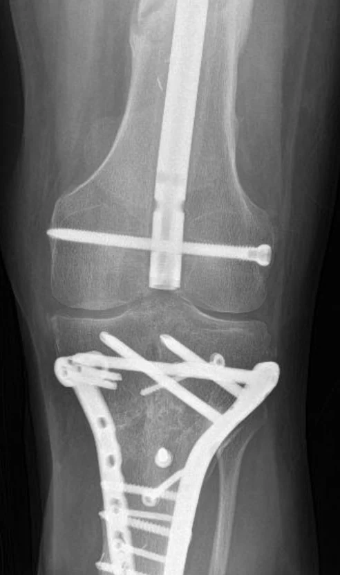
Radiograph of internal fixation with an intramedullary nail and periarticular plate construct
AO Foundation PrinciplesLCP Osteosynthesis

Specialty Intervention 07 · Skeletal Trauma

Trauma & Malunion Reconstruction

Treatment of high-energy intra-articular fractures, open injuries, periarticular disruptions, and established non-unions or malunions. Rigid internal fixation uses anatomically contoured titanium locking compression plates (LCP) and minimally invasive plate osteosynthesis (MIPO).

Clinical Focus

Periarticular & Complex Fractures

Implant Technology

Titanium Locking Compression

Deformity Correction

Corrective Osteotomy

Surgical Protocol

Biological Bone Preservation

British Trauma Fellowship Standards

Precision Matrix

Surgical Modality Comparison

Clinical ParameterConventional JigsPatient-Specific Guides (PSI)Robotic-Navigated (Mako / ROSA)
Alignment Tolerance± 3.0 mm / 3.0°± 1.5 mm / 1.5° 3D template matchedSub-millimeter (± 0.5 mm / 0.5°)
Soft-Tissue TensioningStatic manual tension spacersStatic pre-cut virtual estimateReal-time dynamic sensor graph across ROM
Bone PreservationIntramedullary alignment rodExtramedullary pins with cutting blocksNo canal violation; haptic boundary protection
Pre-Operative 3D ImagingStandard 2D weight-bearing X-raysLow-dose CT with custom 3D printed moldVolumetric CT reconstructed into 3D bone cloud
Recovery Curve6–8 weeks independent gait4–6 weeks structured rehabilitation2–4 weeks with reduced narcotic demand
Ideal CandidatesStraightforward primary deformitiesCases with retained hardwareComplex deformities, active patients, partial knees

Your Orthopaedic Care Pathway

01

Diagnostic Evaluation & Kinematic Scan

Comprehensive 45-minute clinical examination. Weight-bearing radiographs, dynamic MRI, or volumetric low-dose CT alignment scans.

02

Pre-Op 3D Templating & Planning

Sub-millimeter implant sizing, resection angle simulation, and bone-preserve strategy formulated digitally before theatre.

03

Minimally Invasive Precision Surgery

Ultra-clean laminar airflow theatres. Muscle-sparing exposure, computer or robotic guidance, and localized multimodal pain relief.

04

Accelerated Rehab & Long-Term Follow-up

Same-day or Day 1 mobilisation. Structured follow-up at 2 weeks, 6 weeks, 3 months, and 1 year.

Consultant Trauma & Orthopaedic Practice

Schedule Your Consultant Evaluation

Whether you are considering primary joint arthroplasty, seeking a second opinion, or requiring complex tertiary revision surgery, consultations are unhurried and evidence-based.